
It has been almost five years since the outbreak of the COVID-19 pandemic, but the said virus does not seem to be going away any time soon. Further research is still being made on the said virus to develop better vaccines and interventions to prevent its spread, and researchers seem to continue to find new things about the COVID-19 virus.
A recent study from The American Journal of Pathology, published by Elsevier, found an intriguing insight into severe COVID-19-related tissue damage that was never observed in the skin of patients with other types of severe infectious lung diseases or people with only mild or moderate COVID-19. This is in addition to its expected symptoms and respiratory health implications, including breathing difficulties, shortness of breath, and cough.
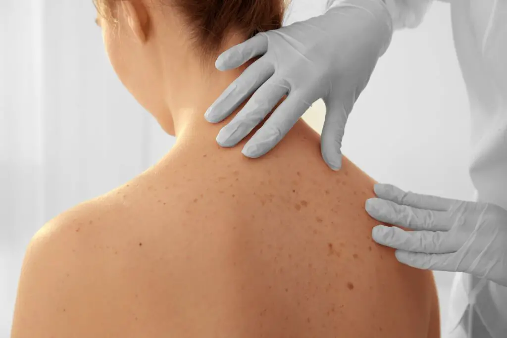
According to the research, this is the first study to present premortem proof of a minimally invasive skin biopsy that can measure tissue damage due to COVID-19 and help identify this blood vessel pathology from other severe respiratory conditions.
Most of those who contract the 2019 coronavirus disease (COVID-19) recover within a few weeks. However, some individuals — even those with moderate forms of the disease — could experience symptoms for a very long time.
One in six persons will experience complications, some of which could be fatal. A condition recognized as cytokine release syndrome or a cytokine storm may be the root of many of these issues. This occurs when an infection prompts your immune system to release an abundance of inflammatory proteins known as cytokines into your bloodstream. They can kill tissue and harm your organs, such as your heart, kidneys, and lungs.
The researchers of the 2022 Elsevier study were the first to realize that acute COVID-19 lung disease was distinct from other severe critical respiratory infections and that the typical pathology was systemic.
The study involved six patients with mild to moderate COVID-19 symptoms, including fever, chills, coughing, or shortness of breath, and 15 patients with COVID-19 who were in intensive care and had their deltoid skin retrieved using a straightforward 4mm punch biopsy technique. The study also included biopsy samples from nine hospitalized patients who passed away before the COVID-19 era and had severe or critical kidney or respiratory disease. Their analysis of the information derived from these biopsies produced very interesting results.
“Patients with severe COVID-19 had clots in small venous and arterial blood vessels in the skin that appears normal and was not found in the skin of patients with other types of severe infectious lung disease, or in individuals with only mild or moderate COVID-19.”
Thirteen of the fifteen patients with severe or critical COVID-19 were found to have microthrombi. Patients with mild to moderate COVID-19, severe respiratory illnesses, or kidney problems in the pre-COVID-19 era had no microthrombi found in their biopsies. COVID-19 respiratory disorders may have these microvascular alterations as a distinguishing feature compared to other acute respiratory disorders.
All six mild to moderate COVID-19 patients had MxA. This antiviral protein can stop SARS-CoV-2 growth, showing that their immune systems were actively battling the virus, as opposed to only two patients with severe to critical illness.
In individuals with severe COVID-19, the microvascular of the skin appeared normal. However, SIN3A, an interferon-induced inflammatory protein, was not present in comparable samples from healthy control people. Increased SN3A expression in skin microvasculature and plasma levels were linked to the severity of the patient’s illness and may be a factor in the cytokine storm for which these patients are known.
The study’s conclusions have clinical ramifications in that they propose that anticoagulants were used in the pre-COVID-19 era in sepsis-associated pneumonia cases to decrease macrovessel thromboembolism. However, the majority of randomized trials to date have not discovered that this treatment is beneficial for hospitalized patients who are critically ill with COVID-19 acute respiratory distress syndrome. These medications might be unable to lessen the microvessel thrombosis associated with SARS-CoV-2 infection.
The nonrandomized referral method was one of the study’s weaknesses. A prospective investigation employing repeated biopsy samples of skin that seems normal is needed before these findings can be proven clinically as relevant predictors of disease development. They stress, however, that a short punch skin biopsy enables tissue-based evaluation of microvascular thrombosis, complement disposition, and MxA and SN3A levels at various periods.
Laurence, J., Nuovo, G., Racine-Brzostek, S. E., Seshadri, M., Elhadad, S., Crowson, A. N., Mulvey, J. J., Harp, J., Ahamed, J., & Magro, C. (2022). Premortem skin biopsy assessing Microthrombi, interferon type I antiviral and regulatory proteins, and complement deposition correlate with coronavirus disease 2019 clinical stage. The American Journal of Pathology, 192(9), 1282–1294. https://doi.org/10.1016/j.ajpath.2022.05.006